Scientific Program

Conference Series Ltd invites all the participants across the globe to attend 15th Euro Congress on Dental & Oral Health Rome, Italy.

Day 3 :

  • Advances in Dentistry | Oral Epidemiology & Community Dentistry | Preventive Dentistry | Dental practice management and Marketing | Dentures | Orthodontics
Location: 1,2,3,4,8,9
Speaker
Biography:

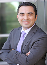
Enrique Cruz has completed his graduation from UNAM Universidad Nacional Autónoma de Mexico in Mexico City where he received his Doctor in Dental Surgery degree in 2000 followed by Training in Oral and Maxillofacial Surgery. After couple years in Private Practice he decided to increase his qualifications by obtaining Dual Specialty Training for 6 years at Indiana University where he received certificates in Periodontics, Orthodontics and oral Facial Genetics, a Master of Science in Dentistry degree and two Minors in Oral Pathology and Life Sciences. His dual training in Periodontics and Orthodontics makes him unique and provides him with the expertise to treat severe cases as well as treating cases of patients who had periodontal problems. He has been involved with dental research and has lectured nationally and internationally presenting his techniques and has been published in the American Journal of Orthodontics and Dentofacial Orthopedics. He is a member of the American Dental Association, American Academy of Periodontology, the American Association of Orthodontist, Southwestern Society of Orthodontics and the Capital Area Dental Society. He is involved continuously with current technologies and techniques to offer his patients state of the art treatment options.

Abstract:

Periodontally accelerated osteogenic orthodontics (PAOO) in an everyday perio-ortho practice Orthodontic patients may benefit from a new surgical technique, Periodontally accelerated osteogenic orthodontics (PAOO). This procedure requires selective alveolar corticotomies and bone grafting a few days after the initiation of orthodontic treatment. This technique may decrease the duration of orthodontic treatment time by more than 50%, reduce root resorption, attenuate relapse and significantly increase alveolar bone support. Rapid orthodontic tooth movement is induced via the surgically-induced regional acceleratory phenomenon (RAP) resulting in an increase in the rate of bone remodeling locally. In this presentation, cases will be reviewed and experiences with PAOO for the past 9 years will be shared.

Anne Charlotte Bas

Paris Sciences et Lettres Paris Dauphine University, France

Title: Role of prices in access in dental cares:A french empirical study
Biography:

Anne Charlotte Bas has completed pursuing in dentistry from Paris Descartes University and a Master’s degree in Health Economics and Public Policies at University Paris Dauphine. She is running for a PhD in Health Economics at PSL Paris Dauphine University on the French Dental Health Market under the direction of Pr. Jerome Wittwer. During this period, they performed 11 communications, several in International Congress, submitted several articles and few are in progress.

Abstract:

In France, dental care prices consist of a public basic fee plus overbilling applied by dentists. Public health insurance proposes very low reimbursement for the basic fee. Extra billings make the most part of costs actually bore by patients. Former studies indeed showed strong out-of pocket and significant inequalities in access to dental care. Dental care concentrated the highest rates of unmet need for financial reasons in France that is 18% of the French population (data ESPS 2012). Dentists decide the overbillings on prosthesis. The general aim of the study is to assess if dentist choices in the pricing process can drive potential financial barriers and social gradient in access to dental care, and how. Our database concerns the years 2006, 2008 and 2010. It is from a match between the French Health, Healthcare and Insurance Survey General Population Survey: ESPS (Enquête Santé et Protection Sociale) and individual consumption from Public Health Insurance data. We explain the unmet need of dental care for financial reason, especially through price. We focus on the inlay-core prosthesis, easy to identify in the data-base and with uniform quality. As a proxy for overbilling distribution in a department, we calculate the median of observed prices in 2010. From an empirical view, we show that unmet needs bring complementary items on access to care. We observed that the individuals living in departments with highest dental fees are 40% more likely to declare to have given up to dental care. The amount of the overbillings on fees, and thus the price which results from, is a barrier of access to dental care.

Biography:

Dr. Mario F. Guiang Jr. Is a graduate of Centro Escolar University Manila Philippines in 1996. Currently he is finishing his Msc. Periodontics in the same university. He underwent series of continuing education in the (University of Illinois college of Dentistry USA ) Europe and Asia. Dr. Guiang underwent further training under the preceptorship of Carl Henley DDS. A renowned cosmetic and neuromuscular dentist Chicago USA. He also underwent training under Dr. Charles Kennedy in Rome Italy a periodontist and Past President of American dental society of Europe. He is the only Filipino lecturer for the Academy of Laser Dentistry USA in 2015 and 2016. His clinical expertise in Neuromuscular Dentistry and Laser Dentistry was chosen among best of the best in the Philippines, USA, Hong Kong, and the United Arab Emirates. He lectures locally and internationally. He is an active member of Academy of Laser Dentistry USA, International association for Orthodontics, International congress of oral implantologist, Philippine Academy of Implant dentistry, Philippine society of Periodontology, Philippine Dental Association, Tarlac Dental Chapter.

Abstract:

Temporomandibular disorders (TMD) happen as a result of problems with the jaw, jaw joint, and surrounding facial nerves and muscles that that control jaw movement. The main cause of this is injury to the jaw, the joint, or the nerves and muscles of the head and neck. This could also be due to the patient’s occlusion not being in the proper position. Transcutaneous electrical nerve stimulation (TENS) currently is a form of electro-analgesia. Hundreds of clinical reports exist concerning the use of TENS for various types of conditions, including myofascial and arthritic pain. It uses electrical currents to stimulate muscle contractions that relieves pain and stiffness while Improving jaw movement. Orthoses is an externally applied device used to modify the structural and functional characteristics of the neuromuscular and skeletal system. They are used to control, guide, and limit joints or body segments for to otherwise correct the shape and/or function of the body, to provide easier movement capacity or reduce pain. Orthoses are also used in denstistry to adjust the patient’s occlusion to a more optimum position, which allows the temporomandibular joint and the surrounding neuromusculatures to be relieved. This poster will present two full mouth reconstruction cases which utilized TENS and orthotics in their treatments. In the first case, TENS and Orthosis was used to increased the occlusion of a patient with severe overbite. While in the second case, TENS and orthosis was used to recapture the occlusion of the patient with posterior open bite. Orthoses used on these patients are tooth colored to preserve the esthetic value of the smile. This poster presents to the dental practioners the importance of restoring a balanced occlusal relationship, and encourages them to incorporate the use of TENS and orthotics in their treatment planning.

Biography:

Noha Ayman Ghallab graduated from Faculty of Oral and Dental Medicine, Cairo University, Egypt, in 1997; and in 2002, she finished her MSc degree and received her PhD in 2005 in Periodontology and Oral Medicine. From 2012 till present, she is an Associate Professor of Oral Medicine and Periodontology at Faculty of Oral and Dental Medicine, Cairo University. She published 12 articles in peer-reviewed international dental journals, supervised more than 20 Master’s and PhD Thesis and is a Peer-reviewer in 4 international dental journals. She attended the International Congress of European Federation of Periodontology “Europerio 8”,
London 2015, with an e-poster.

Abstract:

Introduction: There is low evidence regarding the effect of slow palatal expansion in the treatment of bilateral posterior cross-bite. In adults the treatment had always been directed to surgical maxillary expansion. Therefore, this study aimed at evaluating corticotomyassisted slow palatal expansion using cone beam computed tomography (CBCT). Material & Methods: The study included 6 females, 19-20 years old, with bilateral posterior cross-bite and Class III malocclusion. CBCT was taken pretreatment (T0), immediately post-expansion (T1) and 3 months following the end of orthodontic treatment (T2). Four banded quad-helix appliance assisted with buccal corticotomy and bone allograft was used for expansion. The expansion was activated every 2 months. Fixed orthodontic appliance was performed to continue the treatment of the cases. CBCT was used to measure and record amount of expansion and tipping of premolars and first molars. The bone fenestrations were also recorded as
scores. Data were statistically analyzed.
 
Results: The bilateral posterior cross-bite was treated in 8.5 months. The amount of expansion increased significantly from T0-T1;
5.58±0.77 mm, yet it was non-significant from T1-T2; 1.16±0.34 mm. There was a significant increase in mean tipping angle from
T0-T1 without a significant increase from T1-T2. Regarding buccal bone fenestrations, there was a significant increase from T1-T2
(1.59±1.19, 1.79±1.18 mm, respectively). An inverse correlation was found between bone fenestration and cervical and middle buccal cortical bone thicknesses at T1 and T2.
 
Conclusions: Corticotomy-assisted expansion with quad-helix was an efficient treatment modality of bilateral posterior cross-bite in young adults. Moderate bone fenestration was recorded at the end of expansion.

Biography:

Kallel Ines obtained her Doctors Degree in Dentistry in 2007 and Degree of Specialist in Conservative Dentistry and Endodontics in 2012 from University of Monastir, Tunisia. She started practicing as University Hospital Assistant from 2013 in the Hospital Sahloul Sousse.

Abstract:

Traumatic intrusion is a luxation injury where the tooth is axially displaced into the alveolus. Although, this type of injury is more common in primary teeth, it is considered one of the most severe luxation injuries affecting permanent teeth. Intrusion injuries are often associated with severe damage to the tooth, periodontium and pulpal tissue.
Factors determining treatment choice are stages of root development and intrusion level.The following three methods are only partly evidence based.
• Spontaneous eruption
This is the treatment of choice for permanent teeth with incomplete root formation with minor or moderate intrusion. In teeth with mature root development it is only recommended for teeth with minor intrusion.This treatment seems to lead to fewer healing complications than orthodontic and surgical repositioning. If no movement within a few weeks, initiate orthodontic or surgical repositioning before ankylosis can develop.
• Orthodontic repositioning
This treatment may be preferred for patients coming in for delayed treatment. This treatment method enables repair of marginal bone in the socket along with the slow repositioning of the tooth.
• Surgical repositioning
This treatment technique is preferable in the acute phase. Intrusion with major dislocation of the tooth (more than 7 mm) may be an indication for surgical repositioning. Endodontic treatment can prevent the necrotic pulp from initiating infection-related root resorption. This treatment should be considered in all cases with completed root formation where the chance of pulp revascularization
is unlikely. Endodontic therapy should preferably be initiated within 3-4 weeks post-trauma. A temporary filling with calcium hydroxide is recommended. Through a clinical cases, we will discuss the factors involved in the prognosis of refitting intruders teeth and propose a model treatment plan for this type of dental dislocation.

Gianluca Di Bella

EBSCO Information Services SrL, Italy

Title: Dentistry research through EBSCO online resources

Time : 12:55-13:15

Biography:

Gianluca Di Bella has been working in EBSCO since 2006. He has been Vice President Sales Latin Europe and Israel at EBSCO Information Services since 2010. He has been working for 20 years in the information industry for any institution, library or organization. He is in contact with the most important academic institutions all over Europe and he took part in some health conferences like EAHIL (The European Association for Health Information and Libraries), being EBSCO in the EAHIL network. He has negotiated with the most important biomedical consortia in Europe and he has a big experience in biomedical research databases.

Abstract:

Medical researchers need quick and easy access to the best information available. That is where EBSCO Health can help. EBSCO Health is the leading provider of evidence-based clinical information, healthcare business intelligence, and peer-reviewed medical research tools for the global healthcare community. Content is in the formats that work best for users including medical e-books, medical databases, clinical decision support tools and medical journals. Technology solutions like EDS Discovery Health make it easy for users to access all content from one search box. This is the perfect solution. Users can easily access the medical information they need and purchasers benefit from greater efficiencies, lower cost and increased flexibility. Our goal is to meet the needs of users and maximize return on investment. With Dentistry & Oral Sciences Source, EBSCO Health offers an extensive collection of important full-text journals, many with no embargo. This unique database includes many active full-text sources not available in any other EBSCO databases. Covering top-ranked titles in this discipline, the database includes full-text, indexing and abstracting for hundreds of journals, books, monographs, magazines and trade periodicals, commonly purchased by dental schools and other well-respected institutions. It also offers searchable cited references, linking to the complete text of many of the original works. The database aims to support practitioners, researchers, and advanced dental education. From a statement from the Library.
Journal: “Advanced Search, with its elegantly laid-out tool set for crafting very precise search strategies, gives the user great control
over the whole search process”

Break: Lunch Break@ Hotel Restaurants
Biography:

Nadiah Wasef Ibrahim Al Nahhas, BDS, MSc, Consultant (SCFHS), is also a Consultant in Dental Public Health at the College of Dentistry, King Saud University, Department of Periodontics & Community Dentistry. She is graduated with a degree in Bachelor of Dental Surgery from Cairo University, Cairo, Egypt. She took Master of Public Health from the College of Dentistry, King Saud University, Riyadh, Saudi Arabia in 1998. She became Consultant in Public Health in Saudi
Commission for Health Specialist (SCFHS) in 2006. She was Course Director for three courses in Dental Assistant Diploma Program in College of Dentistry, King Saud University, Riyadh, Saudi Arabia from 2007 to 2012. She is Course Director and Co-Course Director in Preventive and Public Health courses at the College of Dentistry, King Saud University, Riyadh, Saudi Arabia, since 1998 to present.

Abstract:

Introduction: Awareness of gender- or nationality-driven preconceptions can help dentists to have a better interpretation of the dentist-patient relationship. It is even more noteworthy to understand these predilections in Saudi society, where women and men are usually segregated due to religion- and culture-based considerations. This study is one of the first to explore the preferences of patients when selecting a dentist with respect to gender and nationality in the city of Riyadh, Saudi Arabia.
 
Materials & Methods: A total of 445 community residents residing in Riyadh were randomly selected for a cross-sectional study. The participants completed a survey designed to assess which of two factors (gender and/or nationality) were perceived as most relevant in choosing a dentist. Statistical analysis of the data was performed using the SPSS 11.5 software.
 
Results: Female participants did not show any preference for the gender of the dentist; whereas 40% of the male participants preferred a male dentist. Participants also favored male dentists in the fields of oral surgery (78.9%), implants (74.1%), endodontics (67.5%), orthodontics (65.8%) and prosthodontics (64.2%). An exception was noted in pediatric dentistry, for which female dentists were favored by 52.8% of the participants. Additionally, most (66.1%) participants did not have any preference for the nationality of the dentist.
 
Conclusion: Ference for a male dentist but demonstrated no preference for nationality when selecting a dentist.

Hana Omar Al Balbeesi

King Saud University, Saudi Arabia

Title: A Simple Method to Assess Growth Spurt Onset
Biography:

Hana Omar Al Balbeesi has completed her BDS and MSc degrees in Orthodontic Specialty Dental Science from King Saud University/College of Dentistry at Kingdom of Saudi Arabia. She passed the Consultancy Exam from the Saudi Specialized Health Counsel and became an Orthodontic Consultant at the College of Dentistry. She held a position as a Director of Dental Assisting Diploma Program for 4 years. She has published many papers in reputed journals concerning orthodontic diagnosis and treatment planning. She is assisting in teaching of Under-graduate and Post graduate dental students. She participated in many conferences as a speaker.

Abstract:

Since, orthodontists established a strong correlation between tooth developmental stages and skeletal maturational stages; they tried to determine whether dental age assessment is favored over skeletal age maturity assessment. Moreover, they raised many questions to investigate which one of these two methods can be considered a better reliable procedure to predict the future growth potential for conducting appropriate timing for orthodontic intervention, plan proper orthodontic treatment and gain more successful results. Multiple methods were established in the literature to assess dental age, as well as, skeletal age. However, the selected methods in this study were panoramic radiographs and cephalometric radiography, respectively. Previous studies found a strong correlation between third molar development and canine calcification stages in relation to skeletal maturity which was investigated in the literature separately; while others suggested a weak correlation between both with respect to racial dissimilarity. Based on those findings, our present study was aimed to evaluate the validity of dental maturational stages of both canines and third molars, simultaneously, to determine the skeletal maturity, using Nolla’s and Demirjian’s which considered the most valid and easiest methods in estimating the dental age from panoramic radiographs in comparison with skeletal age maturation stages using cervical vertebrae maturational (CVM) method developed by Baccetti and coworkers from cephalometric radiography. Furthermore, to test the accuracy of dental age in estimating skeletal age and whether the dental age can be used as a sole method in determining growth spurt period for planning orthodontic, orthognathic treatment and teeth transplantation for Saudi population.

Biography:

Mohammed Yasser Kharma, BDS, CES, DEA, AEA, is a Lecturer of Oral and Maxillofacial Surgery, Faculty of Dentistry, Aleppo University, Syria; Vice Dean, Faculty of Dentistry, Aleppo University (1997-1999); Consultant of Oral Surgery, Al-Fanateer Hospital, Al-Jubail, KSA 1999-July/2004. He served as Professor of Oral and Maxillofacial Surgery, Faculty of Dentistry, Aleppo University, Syria; Head of Department of Oral Maxillofacial Surgery/Faculty of Dentistry/Aleppo university, 1/9/2010/ until 1/9/2012 and Head of Department of Oral Maxillofacial Surgery at AlFarabi College for dentistry/Jeddah-KSA/ from 1/9/2012/ to till date and Professor in Department of Oral Maxillofacial Surgery at AlFarabi College for Dentistry/Jeddah-KSA to till date.

Abstract:

Objective: To assess the knowledge and attitudes towards Middle East Respiratory Syndrome-Coronavirus (MERS-CoV) among
dental students.
 
Methods: A questionnaire was made according to MOH information and 200 of dental students (Al Farabi College/Jeddah) were
interviewed to evaluate their knowledge about MERS-CoV.
Results: More than half of the dental student 54% interviewed had good knowledge about the etiology, symptoms and treatment
of MERS-CoV. Measurements for infection control and protection were also known 79%. This information collected by students
through College 27%, MOH 25%, Media 24%, Social community 23%, while 17% of interviewed had no idea before.
 
Conclusion: Dental students had good knowledge about MERS-CoV. However more information still must be provided by MOH,
college for medical staff.

Biography:

Muhammad Hasan Hameed has completed his BDS in 2012 from Dow University of Health Sciences, Karachi and has been working as Resident in Aga Khan University, Karachi.

Abstract:

Objectives: Work-related musculoskeletal pain is commonly experienced various health care professionals and dentists are one of them. This study was conducted to assess the prevalence of work-related musculoskeletal disorders (MSDs) among dentists in Karachi and to identify the associated risk factors.
 
Materials & Methods: A cross-sectional descriptive study was conducted among different dental colleges in Karachi during the period of 1 month. Self-administered questionnaire was distributed among 230 dental practitioners for data collection. SPSS version 20.0 was used for statistical analysis. Descriptive statistics was computed and associations of interest were analyzed using chi-square test. Level of significance was kept at ≤0.05.
 
Results: Out of 230 distributed forms, 190 filled survey forms were received. Eight forms were excluded from the study due to incomplete data. The overall prevalence of MSDs in the present study was found to be 75.8%. High prevalence of MSDs was reported in neck (58.8%) followed by lower back (51.6%), shoulders (49.5%), wrists/hands (36.3%) and upper back (34.6%). Statistically significant relationship was found between MSDs and age, specialty, no. of years of practice, awkward posture adaptation and working posture maintained for more than half an hour per patient.
 
Conclusions: Within the limitations of this study, prevalence of MSDs is very high among practicing dentists in Karachi. Most commonly reported sites are neck followed by shoulder, wrists, upper and lower back. The most frequent reasons for MSDs reported were both the lack of rest and static postures maintained for more than half an hour per patient.

Rabia Ali

Aga Khan University Hospital, Pakistan

Title: A Review of Failed Dental Implants at a Teaching Hospital
Biography:

Rabia Ali has completed her graduation from Khyber College of Dentistry Peshawar, Pakistan. She is currently working as a Resident in the specialty of Operative Dentistry in Aga Khan University Hospital, Karachi.

Abstract:

The overall five year success of implants reported in dental literature is 95-97%. Since, the volume of dental implantology is on the rise, thus the failed 3-5% implants constitute a large number of cases. Our study objective was to assess the implants that were placed at a teaching hospital in last five years but later got failed to explore the factors associated with the implant failures. A retrospective charts review was done from June 2010-June 2015. Record of all the implants placed in the hospital was retrieved. The implants that got failed were thoroughly investigated. Variables such as patient age, gender and systemic status, length and diameter of implants, bone grafting, type of definitive prosthesis (crown or bridge or over denture), nature of retention (cement versus screw retained), loading strategy (immediate versus delayed loading) etc. were analyzed. Frequency distribution was computed. Chi square test was applied to compare the association between categorical variables. The unit of analysis was implant. Level of significance was kept at <0.05. Out of 215 implants placed, six implants failed to integrate when assessed for prosthetic phase. One more implant failed after six months of loading. The seven failed cases had following common variables: deficient bone volume in maxilla (or placement of bone graft), non-submerged placement protocol, early loading with prosthesis and presence of diabetes. The five year failure rate of implants was 3.25%. Factors affecting outcome of implant success include atrophic maxilla, premature loading and medical comorbid.

Biography:

Neveen Ahmed graduated in 1999, obtained his BDS degree from the faculty of dentistry at King Abdulaziz University in Jeddah, Saudi Arabia. After working two years at King Fahad General Hospital, He completed his MClinDent program in Pediatiric Dentistry at Queen Mary University in London in 2006. This residency provided his with advanced training in all Pediatric field including treatment of special need children and medically compromise patients. Then Dr. Ahmed moved to Stockholm, Sweden to continue his PhD in Orofacial Pain. He completed his PhD at 2015 from Karoliniska Institutet. Dr. Ahmed is a consultant Pediatric Dentist at Jeddah Specialty Dental Center in Jeddah.

Abstract:

TMJ pain intensity and systemic inflammatory activity in patients with RA are involved in activities of daily living and quality of life impairment. The temporomandibular joint (TMJ) is often and early affected by RA. For example Aliko and coworkers found that 65% of RA patients have TMJ symptoms. The most common clinical finding is TMJ pain, especially on movement or loading. Involvement of the TMJ by RA may, besides pain, cause limitations of jaw function due to restriction of condylar translation. An anterior opening of the bite due to articular cartilage and bone tissue destruction may develop. So, both current TMJ pain intensity and systemic inflammatory activity play roles in the impact of TMJ pain on daily living and quality of life in RA. This implicates that TMJ treatment planning and prognosis estimation in RA patients should not only consider the TMJ pain intensity but also the systemic inflammatory activity.

Break: Break@Foyer

Ziaullah Choudhry

Dow university of health sciences, karachi, pakistan

Title: Bonding of acrylic resin teeth with denture base resin
Biography:

Dr Ziaullah Choudhry is currently working as an Associate Professor in the department of Prosthodontics in Dow University of Health Sciences. Dr Ziaullah did his BDS from University of Karachi and then did his MSc in Prosthetic Dentistry from Eastman Dental Institute London (University College London)

Abstract:

The debonding of acrylic resin teeth with the denture base resin has been related to several different factors of which contamination of the bonding surfaces with wax has been suggested as the major cause. The purpose of this study was to determine the efficient method of wax removal from denture teeth using hot water at different temperatures ranges. Acrylic rods were used as tooth analogues and Raman spectroscopy was used to detect the presence of wax on the surfaces of the specimens. It was found that none of the techniques studied was able to remove all of the wax. Abraded ridge-lap surface showed least wax contamination when dewaxed at 100°C as compared to dewaxed at 85°C and 65°C respectively. The spectrum for each specimen demonstrated peaks at different wavelengths and varying intensities, the spectra demonstrated traces of wax on abraded surfaces showed least wax when dewaxed at 100°C as compared abraded surfaces dewaxed at 65°C and 85° C temperatures respectively.

Biography:

Sadia Tabassum has completed her BDS in 2011 from Army Medical College, Rawalpindi and has been currently working as Resident in Aga Khan University, Karachi.

Abstract:

Objectives: Aim of this study is to compare the depth of cure of two composite materials (SDR & Filtek Bulk-fill) cured at variable
increment depths (2, 4 and 6 mm) and voltages (180 and 200 volts).
 
Materials & Methods: Each sample of the composite material was packed in a mold of 2 mm, 4 mm and 6 mm in a dark room and curing light (quartz-tungsten-halogen) of optimal intensity (above 300 m W/cm2) was exposed for 20 seconds at two different voltages on each specimen. The specimens were then removed out of the mold and the composite on the non-exposed end was then scraped with a flat plastic instrument. The remaining composite represented the set material and was measured using a digital vernier caliper. The reading was divided by half to follow the ISO 4049 method of measuring depth of cure. Student’s t test, factorial design ANOVA and multiple regression analysis were applied. Level of significance kept at 0.01.
 
Results: The mean depth of cure of SDR and Filtek Bulk-fill were 1.93±0.82 and 1.77± 0.65 mm. Lowering the voltage from 220 to 180 volts reduced the depth of Filtek Bulk-fill from 1.87±0.74 to 1.67±0.54 mm whereas; the depth of cure of SDR remained unchanged at 1.93 mm at the two voltages. The adjusted R2 for the depth of cure was 0.93 when the increment thickness, voltage and restorative material were taken together in the regression model.
 
Conclusions: There was no statistically significant difference between SDR and Filtek Bulk-fill for the depth of cure at 2 and 4 mm increments. However, at 6 mm increment, the SDR cured significantly deeper than the Filtek. SDR showed consistent depth of cure at lowered voltage compared to Filtek. Around 91% variation in the depth of cure of these composites materials is explained by increment thickness alone.

  • Technological Tools in Dentistry | Oral Cancer | Oral and Maxillofacial Surgery | Endodontics | Dental Implants and prosthesis Prosthodontics & Periodontics
Location: 2
Biography:

Khaled Ekram, DDS, BDS, PhD, is currently Assistant Professor of Oral and Maxillofacial Radiology at Faculty of Oral and Dental Medicine, Cairo University. His scientific activities include: Senior Consultant of Oral and Maxillofacial Radiology, Member in European Association of Dento-Maxillofacial Radiology, Member in International Association of Dento- Maxillofacial Radiology and Program Instructor for Continuous Education Courses, Faculty of Oral and Dental Medicine, Cairo University, Egypt, 2010-2014.

Abstract:

Cone beam computed tomography (CBCT) is now very widely accepted and is already in use in dental practice all over the world. This presentation will demonstrate how this modality can provide the dentist with useful diagnostic information which can be integrated with other advanced modalities to produce computer generated surgical guides that can allow the operator to surgically apply the predetermined treatment plan for different surgical and prosthetic applications that are difficult or even impossible to obtain by conventional methods. In this presentation, computer guided implant surgery will be discussed in details from A to Z, starting from the CBCT scanning protocol which differs according to the type of support from which the computer generated surgical stent will get its support till the drilling protocol and types of drilling systems and techniques used in these types of surgeries. Also in this presentation, the types of computer generated surgical stents will be highlighted. Surgical stents differs according to the type of support and also according to the drilling technique. Moreover, the precautions that should be made during the surgical protocol will be discussed in details as well as the differences between the conventional surgical protocol and the computer guided methods. Finally, other applications of digital dentistry will be mentioned briefly.

Biography:

Oum keltoum Ennibi is Vice Dean at the School of Medicine Dentistry, Mohammed V University in Rabat, Professor of Higher Education in Periodontology at the School of Medicine Dentistry in Rabat and the Head of Clinical Department of Periodontology. He is Coordinator and Member of several faculty and hospital committees like Research Committee, Continuous and 3rd Cycle Education, Scientific Committee, etc. He is author of several scientific publications in national and international journals and reviewer in national and international journals. He participated in various national and international scientific events. His main research projects include periodontitis especially aggressive periodontitis and gum diseases. He has many research collaborations either with Moroccan teams or international teams (Denmark, Japan and Spain).

Abstract:

Aggressive periodontitis (AgP) are infectious diseases in young subjects harboring an immuno-predisposition to develop the pathology. It is characterized by rapid and severe periodontal destruction in otherwise healthy individuals. Two forms of AgP, localized AgP (LAgP) and generalized AgP (GAgP) have been distinguished based on the number of teeth affected and the distribution of the lesions within the dentition. The presence of Aggregatibacter actinomycetemcomitans (previously named Actinobacillus actinomycetemcomitans) has been correlated with AgP, including LAgP in particular. However, conflicting results for the association between colonization with A. actinomycetemcomitans and LAgP compared to GAgP have been reported for different populations worldwide. Other micro-organisms can be also implicated in the etiology of these entities. In this presentation, we’ll be discussing the clinical features in a Moroccan population and the microbiological aspect of this disease commonly current in Morocco. This discussion will be based on the results of research done so far in Morocco regarding aggressive periodontitis and presence of A. actinomycetemcomitans. Regarding the destructive potential of these diseases, patients lose their teeth in relatively early age and they may also present severe teeth migration; thus, dental and esthetic management is often difficult and costs a lot. Possible treatment modalities will be discussed regarding the bacterial etiology; some inter disciplinary management approaches will be presented. Finally, we will emphasize the role of the dentist on early screening and management of this periodontal destructive disease in young subjects knowing that a familial aspect of aggressive periodontitis has been largely highlighted by many studies.

Break: Break@ Foyer
Biography:

Ghada Gamal Adayil has completed her Bachelor’s in Dental Medicine in 2012-excellent degree with honor from Faculty of Oral and Dental Medicine, Cairo University. She was assigned as a Dentist at Ministry of Health for one year then as Internal Resident Doctor at Periodontology Department at Faculty of Oral and Dental Medicine, Cairo University till now. She is a currently a Master’s degree student.

Abstract:

Plasma cell gingivitis (PSG) is a rare inflammatory condition manifested as erythematous enlarged gingiva. It may be associated with cheilitis and/or glossitis. A 47-year-old woman presented to Department of Periodontology, Faculty of Oral and Dental Medicine Cairo University, complaining from eating difficulties and teeth mobility. Clinical examination revealed severe gingival enlargement affecting the whole width of attached gingiva from both facial and lingual aspects. The enlarged gingiva was erythematous and friable with nodular appearance. Periodontal examination showed generalized advanced degree of periodontitis with the anterior teeth affected more than the posterior one. The affected anterior teeth were covered by the enlarged gingiva, nearly without bony support or with minute support less than the apical one third of the root length, with all showing pathological migration and grade three mobility. Lips were enlarged, dry, atrophic and fissured with angular cheilitis. There were no relevant findings in her medical history and she was not taking any regular medication. Laboratory investigations confirmed the absence of relevant systemic disorders. Conventional periodontal therapy was performed then extraction of all anterior teeth and surgical excision of enlarged gingiva were done. Histological examination showed that both epithelium and connective tissue were infiltrated with inflammatory cells. The epithelium showed spongiosis and underlying connective tissues contained dense infiltration of plasma cells. Plasma cell gingivitis was described in the literature in association with aggressive periodontitis. This case represents plasma cell gingivitis condition of unknown cause associated with generalized chronic periodontitis. We can describe the case as Plasma cell Periodontitis (PCP) instead.

Biography:

Nabiel ahmad alghazali: Dr. Alghazali N is working as consultant prosthodontics at Riyadh, Saudi Arabia.He finished his diploma and PhD.

Abstract:

Different protocols have been used in practice to produce an accurate impression for full mouth implant supported prostheses. This including: Different impression materials; different impression technique; splinting or non-splinting of impression cops; different splinting materials and different splinting protocols. In this review paper, literature has been reviewed to finalize the ideal and most predictable impression technique to be used to give and accurate and well-fitting final prostheses in case of full mouth implant supported prostheses. A clinical case also was presented here to demonstrate a step by step protocol for this purpose.

Biography:

Mohammed Hussein Al Bodbaij, BDS (KSU), MSc OMFS (UCL), MFD RCSI, got his Bachelor’s degree of Dental Surgery (BDS) in 1997 from King Saud University at Riyadh. He joined Ministry of Health at KSA soon after internship year to work in Primary Health Care Center. After 2 years, he transferred to Oral & Maxillofacial Surgery Department at King Fahad Hofuf Hospital (KFHH) to work as a Resident. Two years later, he attended MSc OMFS course at Eastman Dental Institute at University College London where he got the degree in 2005. After that, he returned back to KFHH to work as a Specialist. Since 2007, he has worked as an Acting Consultant. In 2013, he got the Fellowship of MFD RCSI, Ireland. During his career, he gave many local and international speeches and had few publications. He is a member of Saudi Dental Society and member of Saudi Society of Oral & Maxillofacial. At present, he is the Head of Oral & Maxillofacial Surgery Department at King Fahad hospital-Hofuf.

Abstract:

Central giant cell granuloma (CGCG) is a benign lesion; CGCG occurs mainly in children and young adults with more than 60% of all cases occurring before the age of 30 years and female to male ratio of 2:1. The mandibular/maxillary ratio is from 2:1 to 3:1. Surgery is the traditional treatment of CGCG. Calcitonin and intralesional steroid were used with good results. In this case report, a 14 years old Saudi girl presented with a hard swelling of left side of the mandible with few months duration. Investigations including blood tests, radiographs and biopsy were done which confirmed the diagnosed of CGCG. Lesion has been treated using 6 weekly intralesional injections of steroid which gave very good result. Patient has been followed for more than 16 months with radiographic evidence of defect refill with bone and no sign of recurrence.

Biography:

Dr. Sheetal Rao is currently pursuing her Master Degree Program in the Department of Conservative Dentistry and Endodontics, Manipal University, India. Her areas of specialty include disinfection of root canal systems, laser dentistry and bioactive materials. She is actively involved in a number of ongoing research work and has contributed to various publications. She also mentors undergraduate students in their theory and practical work. She enthusiastically participates in numerous pro bono activities throughout the year.

Abstract:

It has been widely considered that, endodontically treated teeth are more susceptible to fracture than vital teeth. This is because, endodontic procedures results in structural changes of root canal dentin that makes the tooth prone for vertical root fracture (VRF). Therefore, the residual tooth structure should be reinforced via root canal filling materials to prevent VRF. Recently, it has been suggested that mineral trioxide aggregate (MTA) was able to produce a tight seal with the root dentin walls that was superior to many other existing materials. However, the ability of MTA to strengthen the tooth structure has been studied with controversial results. White et al. demonstrated weakening of dentinal structure in short term and attributed this effect to the structural alteration of proteins caused by the alkalinity of MTA. Andreasen et al. did not find a significant increase in fracture strength when immature sheep teeth were filled with MTA and compared with control (without filling) teeth. However, the strengthening effect of MTA has been shown in other studies. OrthoMTA (BioMTA, Seoul, Korea) is a newly developed calcium silicate cement which is known to have shorter setting times (5 h 30min) and less heavy metal content. According to the manufacturer, OrthoMTA prevents microleakage by forming an interfacing layer of hydroxyapatite between the OrthoMTA and the canal wall. Furthermore, it exhibits a bioactive characteristic; it releases calcium ions through the apical foramen, and the released calcium ions help to induce regeneration of the apical periodontium. Yoo et al. has reported that, the OrthoMTA-PBS paste has the potential antibacterial effect in infected root canals. Recently, OrthoMTA III (BioMTA, Seoul, Korea) has been marketed in which, Zirconium oxide has been used a radiopacifier to prevent discoloration compared to the older version of OrthoMTA which had bismuth oxide as radiopacifier. Till date, there was no study to evaluate the efficiency of OrthoMTA III in reinforcing the endodontically treated teeth when used as an obturating material. Hence, the aim of this study was to evaluate the influence of ProRoot MTA and Ortho MTA III as an obturating material on the fracture resistance of endodontically treated. Group I-control (no root canal filling); Group II- ProRoot MTA; Group IIIOrthoMTA III. In group II and III, MTA powder was mixed with distilled water following the manufacturer’s instructions. MTA was then inserted into the root canals using MTA carrier and condensed incrementally using pluggers leaving the coronal 2 mm of the canal unfilled. A glass slab was used to prevent the extrusion of MTA from the apex. All the teeth were radiographed to verify the homogeneity of the fillings. Coronal access cavities were sealed with glass ionomer cement (Fuji II) and the specimens were stored at 37 o C and 100% relative humidity for 48 h. Periodontal ligament (PDL) simulation was performed by the modified method of Soares et al. The roots were coated with a thin layer of moulding wax of 0.3-0.5mm thickness up to 2.0 mm below the cementoenamel junction. The roots were then embedded in polystyrene resin using polyvinyl chloride cylinders (PVC). After resin polymerization, the teeth were removed from the PVC cylinders, and the wax were removed from root surface and resin cylinder using hot water. The resin cylinders were then filled with a polyether impression material using a syringe. The roots were then re-inserted into their respective cylinder ‘sockets’ and excess impression material was removed with a scalpel blade.

Biography:

Anum Aijaz has completed her BDS in 2011 December from Liaquat University of Medical and Health Sciences, Jamshoro Hyderabad and has been currently working as a Dental/Medical Officer in Aga Khan University, Karachi.

Abstract:

Background: It is essential to properly manage the gingival tissues for obtaining accurate impressions when restoring tooth structure which is in close proximity to the gingiva and for fabricating fixed restorations, particularly when the finish line is near the gingival sulcus. The retraction of the gingiva creates sufficient lateral space between the tooth preparation finish line and the gingival tissue with minimum trauma, to allow for the injection of adequate bulk of impression material into the gingival crevice. There are various gingival retraction methods to choose from including mechanical, chemo-mechanical and surgical methods.
 
Objectives: The purpose of this systematic review was to see which gingival retraction method was most effective in terms of amount of gingival retraction achieved and minimal trauma to the gingival tissue as assessed by clinical parameters. Methods: In addition to search engines like PubMed, CINAHL plus (Ebsco) and COCHRANE, hand search was also used to search articles related to our review question using the key terms and different permutations: Gingival retraction* AND displacement method* OR technique* OR agents OR material* OR medicament* OR apparatus. The systematic review was registered on Prospero registration no CRD420140133335. Our initial search results yielded 142 articles which were narrowed down to final 7 articles after a strict eligibility of including clinical trials on gingival retraction methods with the amount of tooth structure gained and assessment of clinical parameters GI, PI, PD, AL, BOP, etc. as the outcomes, conducted on human permanent teeth only.
 
Results: The total number of teeth assessed in all 7 studies was 336. The surgical site was mentioned in 6/7 studies whereas type of jaw was mentioned in only 3/7 studies. Most commonly used gingival retraction method was mechanical. 3/7 studies reported the amount of gingival retraction achieved whereas 4/7 studies assessed the clinical parameters.
 
Conclusions: There are just 7 studies (trials and experimental) available on the retraction methods. The studies were heterogeneous in terms of reporting the outcomes (amount of gingival retraction achieved). No single method (mechanical/chemo-mechanical/ surgical) appeared superior to the other. Clinical parameters such as PI, GI, PD, AL, BOP, etc. were not significantly affected with gingival retraction.

Muhammad Adeel

Dow University of Health Sciences, Pakistan

Title: Comparison of removal potency of different intracanal medicaments
Biography:

Dr. Muhammad Adeel Ahmed is currently working as an Assistant Professor in the Department of Endodontics at Dow University of Health Sciences. He has completed his Membership from Royal College of Surgeons of Edinburgh (United Kingdom) in 2011 and four year Fellowship in Endodontics from Pakistan College and Physicians and Surgeons in 2013. At present, he is enrolled in indigenous PhD program at Dow University of Health Sciences. He has Seven publications in peer reviewed journals. He is also Dental Postgraduate Programs Co Ordinator and Supervisor for Master’s Program at Dow University of Health Sciences.

Abstract:

Aim: To compare the removal potency of calcium hydroxide [Ca(OH)2], triple antibiotic paste (TAP) and doxypaste using manual irrigation with sodium hypochlorite (NaOCl). Design In vitro experimental study. Place and Duration of the study The study was conducted at DIDC and DRIBBS, Dow University of Health Sciences and Department of Material Sciences, NED University of Engineering and Technology in December 2015. Methodology 30 extracted single and multirooted teeth having 45 canals were prepared using k files upto MAF. The canals were divided into 3 groups (15 canals in each group) receiving Ca(OH)2, triple antibiotic paste and doxypaste respectively. After storing these teeth in incubator at a temperature of 37o C with 95% humidity and darkness for a period of 15 days, these medicaments were removed by manual filing followed by irrigation with side vented needle at 1mm from working length using sodium hypochlorite. All samples were then observed under a stereomicroscope after sectioning. The results were analyzed on SPSS Version: 16 using kruskal-Wallis test (P-value 0.05).

Result: Remnants of medicaments were found in all three of the experimental groups with calcium hydroxide being associated with highest amount of residues (33.3%) which was more than 2 times greater than triple antibiotic paste (11.6%) and 6 times greater than doxypaste (5.72%).

Conclusion: Failure in removing these medicaments completely from the canal walls may adversely effect the endodontic treatment outcome by compromising the efficacy of the sealer.

Break: Lunch Break@ Hotel Restaurants
Biography:

Dr. shaheen ahmed is currently working as an Assistant prof in oral and maxillofacial surgery department in dow university of health sciences. He have done my Fellowship from college of physician and surgeon Pakistan in 2011 in oral and maxillofacial surgery. He also the supervisor of MDS program in dow university of health sciences having 6 postgraduate trainee. He had published 4 articles and two case reports and also perusing PhD from Dow university.

Abstract:

This descriptive case series study assessed the spectrum of maxillofacial injuries seen at the Emergency Department of Abbasi Shaheed Hospital, Karachi, Pakistan from March 2009 to February 2010. A total of 1295 patients with maxillofacial injuries were included in the study, of whom 875 (67.6%) were male and 420 (32.4%) female. Injuries were most common 340 (26.25%) in first decade. The most common cause was road traffic collision (611) patients and fall was second most common cause (415) patients. The mandible. was the most common bone fractured (309) patients, followed by dentoalveolar (142) patients. There were 73 patients with fractured maxilla. Isolated soft tissue injuries occurred in 578 patients most commonly occurring in the lower third of the face. Most of the maxillofacial injuries were received between 1500 and 2100 hrs, (665) and Saturdays were the busiest days with (242) patients. The collection of data over the one from emergency department regarding the maxillofacial fractures including soft tissues injuries helps us how to manage on early basis and also may guide us regarding the time and day of the week which is busiest time of
the emergency department.

Biography:

Muhammad Ehsan ul Haq, BDS, FCPS is assistant Professor at Akhtar Saeed Medical & Dental College, Lahore, Pakistan in Oral & Maxillofacial Surgery Department.

Abstract:

Background: The oro-facial region including the jaw bones and related tissues can be the site of a multitude of neoplastic conditions. Rare odontogenic/ non odontogenic tumors have variable pattern, clinical behavior, radiographic appearances as well as prognosis. Surgical resection with safe margins is needed in most of the cases. Odontogenic myxoma, fibrosarcoma, ewings sarcomas, orbital osteomas were quite rare tumors of the maxillofacial region. The treatment modalities were usually surgery with or without radio & chemotherapy. It is envisaged that results presented here will add to the depth of information on facial tumors in this region and in Pakistan at large.
 
Aim: Pattern and treatment outcomes of rare tumors of the oral and maxillofacial region.
 
Methods: Patients of all age and sex presented at the Department of Oral and Maxillofacial Surgery, KEMU/ Mayo Hospital Lahore with tumors affecting the Oro-facial region from March 2010 to March 2016 were included.
 
Results: Total 106 cases were included. Among uncommon benign and aggressive tumors (n=69): odontogenic myxoma n=11. CEOT n=12, ossifying fibroma n=15, AOT n=09, nasopharyngeal angiofibroma n=13, Ethmoidal osteoma n=04, giant cell tumour n= 5 cases were included. Among rare malignant tumours (n=37): fibrosarcoma n=09, Ameloblastic carcinoma n=10, adenoid cystic carcinoma n=09, mandibular mucoepidermoid carcinoma n=4, ewing’s sarcoma n=05, melanoma n=04 cases.
 
Conclusions: Benign tumors more common in mandible except AOT which is common in maxillary canine region. Surgical management of tumors in the maxillofacial region is complex and challenging. Large mandibular defects can be bridged with reconstruction plates only.
 

Ahmed el-hoshy

Cairo University, Egypt

Title: Dentin hypersensitivity
Biography:

Ahmed el-hoshy is working as an Associate Professor of Conservative Dentistry, Cairo University and Future University, Esthetic course Director in Continuous Education Program in Future University. He serves as Post-graduate course Director for Esthetic Dentistry in Future University and Key Opinion Leader with Philips Discuss Dental for tooth bleaching products. He is Scientific Advisor for k59 ceramic bur, Komet, Brasseler.

Abstract:

Dentinal hypersensitivity is a very common clinical condition usually associated with much routine dental treatment in our daily practice such as scaling and bleaching. It can affect patients of any age group in both arches. This lecture concisely reviews the etiology, mechanism and clinical management. Treatment should start with an accurate diagnosis. Differential diagnosis should be made and all other probable causes should be excluded. We are focusing to help the operators to solve this problem in a straight and easy way.

Break: Break@ Foyer
Biography:

Sheikh Bilal Badar has completed his BDS in 2012 from Dow University of Health Sciences, Karachi. He has published two papers in reputed journals and has been currently working as Resident in Aga Khan University, Karachi.

Abstract:

Introduction: Various methods are described in literature for determining accurate age, one of which is coronal pulp index by using
dental radiograph.
 
Aim: The aim of present study is to determine the correlation between chronological age and coronal pulp cavity index in a sample
of Pakistani population.
 
Material & Method: Retrospective charts review was conducted in AKUH dental clinics at two months period in which a total of 332 teeth (178 females and 154 males) of Pakistani individuals were analyzed on panoramic radiographs using non-probability convenient sampling technique. Coronal pulp chamber heights and crown heights were measured in unrestored mandibular right and left first and second premolars, and first and second molars. These measurements were then used to calculate the tooth coronal pulp cavity index (TCI). Data was analyzed by using SPSS version 19. Independent sample t-test was used to assess bilateral symmetry of TCI for premolars and molars. Pearson correlation was used to assess association between chronological age and TCI.
 
Result: The correlation coefficient between chronological age and tooth coronal index was found to be -0.13. The correlation coefficient in males ranges from -0.03 (for tooth#44) to -0.47 (for tooth#47) whereas in females, the correlation coefficient ranges from -0.02(for teeth#44) to 0.41 (for tooth#46). However, tooth#47 in males showed some significant degree of negative correlation
(r=-0.47) and tooth#36 in females showed significant degree of negative correlation (r=-0.51).
 
Conclusion: This study shows that there is very weak association between age and TCI of any tooth in Pakistani population so coronal pulp index cannot be accurately used for age estimation of Pakistani population. Other methods should be used for accurate age estimation.

Biography:

Belkacem Chebil Raouaa has completed her PhD at the age of 25 years from Monastir University and postdoctoral studies from the same university. She is now an assist professor in oral medicie oral surgery. She has published some papers in reputed journals.

Abstract:

Objective: Low-level laser therapy (LLLT) is a treatment commonly used in oral surgery for biostimulation. The aim of this study was to compare the effects of LLLT and corticosteroids on postoperative pain, trismus and edema following surgical removal of mandibular third molars.
 
Background Data: LLLT and corticosteroids have been effective in reducing pain, trismus and edema; thus improving the quality of patients’ lives.
 
Patients and Methods: Sixty patients who were to undergo surgical removal of their lower third molars were studied. They were randomly assigned to two groups of thirty patients each. A diode laser device (Whitening laze II) with a continuous wavelength of 808 nm and a maximal output power of 100 mW was used. Patients in the LLLT group received 3.3 J (fluence of 120 J/cm2) intraorally at the operation site and the same dose extraorally. Patients in the corticosteroids group received postoperative parenteral injection of Dexamethasone. Pain, interincisal opening and facial swelling were evaluated on the third and seventh postoperative days.
 
Results: The levels of pain, trismus and facial swelling at the third and seventh postoperative days were lower in the corticosteroids side than in the LLLT side, though without statistically significant differences except for edema at seventh postoperative day.
 
Conclusion: This study demonstrates that there is no significant difference between administration of corticosteroids and LLLT for the reduction of postoperative pain and trismus but dexamethasone was more effective to reduce swelling on seventh postoperative day.

Biography:

Elnaz Safdarian is senior at Zanjan university of medical sciences.

Abstract:

Introduction: Depression, anxiety and stress prevalence is important problem among dental students. Due to psychiatric side effects among students, this study was done for evaluating the amount of depression, anxiety and stress among Zanjan dental students in academic year of 2015-2016(Iran).
 
Material and Methods: This study with designing of cross sectional-description was done on 160 people of dental students. Samples selection were accidentally and information were completed by questionnaires. In this research questionnaire DASS21 was used for investigating amount of depression, anxiety and stress and questionnaires DESQ was used for investigating stress source factors. Results were analyzed statistically by t-test and chi-square.
 
Results: This study showed 31.5%, 40.3% and 41.6% of dental students have faced depression, anxiety and stress respectively with various intensities. Depression, anxiety and stress levels were not significantly different statistically in two genders and also students with various residence locations (p>0.05). Also depression and stress was significantly different statistically by taking into consideration of academic year (p<0.05). The most important stress source factors from students’ point of view were as follows:
• In educational field was fear of failing (57%)
• In clinical field was responsibilities for comprehensive patient care (36.2%)
• In outside of university was living far from family (37.5%)
Conclusion: According to mentioned depression, anxiety and stress prevalence and of course existing stress source factors, it is recommended that authorities endeavor to decrease these factors and prepare a better educational atmosphere for students.

Monika Balkandzhieva

Medical University of Sofia, Sofia Bulgaria

Title: Tobacco smoking and chewing. Oral cancer.
Biography:

Monika Balkandzhieva are 4th class of dentistry in Faculty of Dental Medicine of Sofia of Medical University of Sofia, Bulgaria. Alper Yilmaz is Chairman of the General Meeting of the student council of Medical University of Sofia.

Abstract:

It is now accepted that there is an etiological relationship between the smoking of tobacco and oral carcinoma regardless of the type of tobacco and method of consumption. Pipe and cigar smoking have been linked with carcinoma of the lip for many years, and the evidence linking cigarette smoking with intraoral carcinoma is now firmly established. Studies have particularly incriminated heavy cigarette smoker and have shown that those smoking 40 to mote cigarettes per day have a significantly increased risk of oral cancer, ranging from about 10 to 20 times that of non-smokers in different series. The type of tobacco, curing methods, and methods of smoking may also influence the relative risk of oral cancer. For example, the high incidence of oral cancer in India is likely to be due, in part, to widespread smoking of bides and the habit of reverse smoking, a habit that is particularly common in women. Reverse smoking is also practiced in various other countries, for example Colombia, and is associated particularly with cancer of the palate, one of the rarest sites for oral cancer in other groups. It has been reported that the relative risk of oral cancer for reverse smokers is over 40 times that of non-smokers. Smokeless tobacco: Snuff is a finely ground or powdered tobacco which may be inhaled dry or used moist in snuff-dripping by placing a pinch of snuff between the gum and the cheek or upper lip. Report from South-Eastern USA, where snuff-dripping is prevalent, and from Sweden indicate that the habit is associated with a significantly increased risk for carcinoma of the gingiva and buccal mucosa.Tobacco chewing was relatively common in the United Kingdom in the early part of 20th century, particularly in occupations such as mining where smoking was environmentally dangerous because of the possibility of explosion.

  • Extended Networking Session and Lunch@ Foyer