Scientific Program

Conference Series Ltd invites all the participants across the globe to attend 25th Euro Congress and Expo on Dental & Oral Health Budapest, Hungary.

Day :

  • Orthodontics| Endodontics | Dental and Oral Health | Oral and Maxillofacial Surgery | Laser Dentistry
Location: Budapest, Hungary
Speaker

Chair

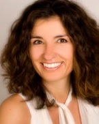
Eleni Stil

CEO, Style for your smile, Germany
Speaker

Co-Chair

Randa Zein

Laser dentistry, University of Genoa, Italy

Speaker
Biography:

Altukroni, Abdulbadea Abdulrahman M has completed his bachelor at the age of 24 years from October 6 University and his work now in ministry of health in Saudi as a resident dentist.

Abstract:

The aim of this study is to prove the effeteness of this devise that have been developing. The ultrasonic excavator will be easily used to penetrate in caries and defective dentine which will decrease traumatic injury through connecting ultrasonic wave to excavator tip.
Objectives: Remove the caries and defective dentine with less trauma, and laceration to the pulp tissue, also, to reduce heat generation, vibration, noise and the hand pieces pressure applied on the tooth that come from air driven by using ultrasonic excavator According to Laird and Walmsley in the solution for all problems that are mentioned above.(1991) it is possible to use ultrasound without the resource of analgesia. And according to A.g.Antonio, L.g.primo and L.c.maia (case report 2005) the ultrasonic technique is the only one able to remove carious dentine without the formation of a smear layer and the consequent obstruction of dentinal tubules, and in same case report he said about using the ultrasonic technique for caries removal it is provides more safety during the treatment and offering comfort to the patient. And according to Ming-shu l (2013 animal study) if we make perforation on a tooth by ultrasonic device is better than making it by air driven handpieces based on the bleeding level, histologically and time to become a necrotic pulp after perforated. As conculsion: According to my experience when I used the ultrasonic excavator to excavate the caries and defective dentine for two molars extracted teeth, it proved to me the devise can removed a caries and defective dentine. But the device under development and there is a trial version of it and also I have patent application from Saudi patent office, number 116380118.

Speaker
Biography:

Dina A Morsy has received her Master’s Degree in 2013 from Faculty of Oral and Dental Medicine, Cairo University, Egypt. She is a PhD student in Faculty of Oral and Dental Medicine, Cairo University, Egypt. She is working as an Assistant Lecturer in Endodontic Department, Faculty of Oral and Dental Medicine, Cairo
University, Egypt.

Abstract:

The main goal of root canal treatment is total eradication of the microorganisms to attain bacteria-free environment both in the tooth and at the apex, including the periodontal tissue and the surrounding apical bone to favor the environment for healing without the need of periapical surgery in some resistant cases. Recently the use of lasers has gained an increased interest in the endodontic field due to its bactericidal effect and bio-stimulation effect. In this case report, five cases diagnosed with necrotic pulp and periapical lesion were selected. A CBCT scan for each patient to verify the size (5-10 mm) and bone density of the periapical lesion preoperatively. Microbiological samples were taken after accessing the root canal, after conventional root canal treatment and after canal irradiation with diode laser (980 nm) coupled with the optical fiber 200 μm to assess the bacterial count after each procedure. A second CBCT scan was taken for each patient at 6 months follow up to evaluate the healing process. Complete eradication of the bacteria after canal irradiation with diode laser was obtained. The evaluation of the CBCT scan showed decrease in the periapical lesion size and increase in its density after the 6 months follow up. It can be concluded that laser therapy may be used as an adjunct to conventional endodontic therapy to improve the treatment quality of infections of the oral cavity. This may lead to save the patients from the invasive surgical intervention.

Speaker
Biography:

Randa Zein MD, DSS , PHD candidate . She was completed her MSc laser dentistry university of Genoa Italy, Restorative and Esthetic specialist Toulouse university –France. She is a Head of the laser division in the international unions of universities. She is doing Private practice at Lebanon Beirut – Hamra – Main Street. She had many article on Laser Dentistry and she is Authors and co- author of several articles on laser dentistry

Abstract:

Practical Application Of Lasers In Every Day Dentistry
 
Background: In the past decades the concept of “Laser applications in Dentistry“has been established. This new concept implies several changes to widely adopted “cut and suture” and radical strategies when managing the caries and gingival procedures. New generation of Erbium and diode lasers have been introduced to dental clinics with very high success and patient acceptance rates. What can’t not be solved using the traditional treatment, laser can be a good choice to resolve it.
In this lecture we will discuss the details of lasers application of hard and soft tissue lasers
1. What are the advantage of using laser over the traditional treatment
2. Hard tissue applications of lasers in dentistry
3. Soft tissue application of lasers in dentistry
4. Clinical cases presentation
5. Low level laser therapy from science to practice
 
Methods: A recent review about the use of lasers in dentistry was conducted .
 
Results: Dentistry today is evolving tremendusly and the use of lasers in dentistry is a double –edged sword : if it is properly used, laser can be an initial and/or additional tool in our daily practice , whereas if it is misused it can lead to catastrophic results.
 
Introduction To Laser Dentistry
 
The introduction of laser in dentistry, in the 1960s, by Miaman, led to a continuous research in the various applications of lasers in dental practice. There are two scenarios, on the one hand, there are hard lasers, such as, Carbon dioxide (CO2), Neodymium Yttrium Aluminum Garnet (Nd: YAG), and Er:YAG(Cr), which offer both hard tissue and soft tissue applications, but have some limitations: whereas, on the other hand there are cold or soft lasers, based on the semiconductor diode devices, which are compact, low-cost devices . Laser use in dentistry has expanded and improved some treatment options for clinicians who have adopted this technology
 
The objective of this lecture is to provide an overview on :
1-How do lasers work in dentistry
2-What are the common components of all lasers devices
3-Laser physics and laser tissue interaction
3-The advantages of using this technology in private clinic
4-The kinds of devices that are the best to use for every dental specialty
 
Methods: A recent review about the use of lasers in dentistry was conducted .
 
Results: Dentistry today is evolving tremendusly and the use of lasers in dentistry is a double –edged sword : if it is properly used, laser can be an initial and/or additional tool in our daily practice , whereas if it is misused it can lead to catastrophic result.


Magda Eline Guerrart Portugal

Universidade Federal do Parana , Brazil

Title: Xerostomia Related To Medication Use In The Elderly

Time : 14:00-14:30
Speaker
Biography:

Magda E G Portugal is a Ph.D student in the Internal Medicine post-graduation programe at Universidade Federal do Parana in Brazil. She is a dental surgeon and has been working in virology research, epidemiology and teaching for five years. Currently, her research is related to know the frequency of HPV infection and its association with head and neck tumors, identifying the HPV genotypes, the expression of viral genes and cellular proteins. Her expertise in Public Health Education, specially with elderly patients came after some years of experience in research and teaching both in hospitals and education institutions.

Abstract:

In Brazil, 8.6% of the total population is 60 years old and most of them consume 60% of the national production of medicines. Due to the mouth-related (oral) side effects of medications and its impact on public health, the aim of this study was to analyze the association between salivary flow and the use of medications in the elderly, as well as to enquire if patients who use medicines were told about the possible side effects in the oral cavity. Methodology: This research was a case study (n = 30) of people aged over 60 that attended the dental clinic of Herrero School of Dentistry, approved by the ethics committee of the institution. Unstimulated saliva samples were collected and data were obtained through a questionnaire, after application of IC. Salivary volume flow and pH were assessed, and data were subjected to statistical analysis with p = 0.05 significance level. Findings: None of the patients in this study had ever received any professional information about possible oral side effects of medication. The results showed that the salivary flow of 59.37% of the sample was between 0 and 0.25 ml / l and that 68.75% had a pH between 6.1 and 7. The use of some medication was reported by 78.1% of the sample and the all the 14 pharmacological groups indicated were significantly associated with changes in the oral cavity such as dry mouth, difficulty swallowing, chewing and difficulty swallowing solid foods.

Speaker
Biography:

Dr. Dustin Pfundheller is the world's most traveled surgeon and dentist. He compares the way dentistry is performed throughout the world - including Asia, Mideast, Caribbean, Africa, Pacific, Europe, Isolated/remote places, USA, and South America. Dr. Dustin Pfundheller is a general dentist who has been successfully performing braces for several years. He will cover the basics of bonding brackets, case selection, archwire sequence, leveling and finishing. Discussions will be done on how to recognize potential complications and when to refer to an orthodontic specialist. Dr. Dustin Pfundheller w ill cover the skills and knowledge needed to start using implants in your daily practice. Participants will understand how to treatment plan, place and restore dental implants in both the anterior and posterior
regions.

Abstract:

The lecture will cover the skills and knowledge needed to start using implants in your daily practice. The participants will learn indications and contraindications to dental implant related treatment. They will understand how to treatment plan, place and restore dental implants in both the anterior and posterior regions.

Speaker
Biography:

Dr Young Gun Education Details: Mar 2002 ~ Feb 2008 : DDS, predental course and dentistry, College of Dentistry, Yonsei university, Seoul, Republic of Korea Mar 2010 ~ Feb 2013 : Residency program, Department of Orofacial pain and Oral medicine, College of Dentistry, Yonsei university, Seoul, Republic of Korea Apr 2013 ~ Apr 2016 : as a Army surgeon in dental surgery, Republic of Korea Army May 2016 ~ Present : Fellowship, Department of Orofacial pain and Oral medicine,
College of Dentistry, Yonsei university, Seoul, Republic of Korea.

Abstract:

The aim of this study was to simplify the optimal temporal areas for safe and reproducible approach for BoNT injections into the temporalis muscle(TM) by carrying out detailed dissections and measurements of the structures in the temporal area, and to virtually represent a topographic mapping of postural relations among the major anatomical structures such as superficial temporal artery, middle temporal vein and temporal branch of the facial nerve in the temporalis muscle of the temporal area. Nineteen sides of TM from 10 embalmed Korean cadavers were used in this study. The lateral canthus of the eye and tragus were set as landmarks to establish the reference line of this study. The topographies of the superficial temporal artery, middle temporal vein, temporalis tendon, and the temporalis muscle were evaluated. On the disclosed boundary of the muscle, we can visualize an imaginary, rectangular TM in the temporal area. The surface of TM can be divided into 9 equally sized rectangular areas. The topography of studied anatomical structures in these nine compartments was observed and measured from the superficial to deep layers. After drawing the muscle boundaries, they were divided into nine compartments in order to simplify the relationship between the soft-tissue landmarks and the anatomical structures, and to more facilitate the description of appropriate injection sites. The relative positional ratios between the anatomical structures were constant in all of the specimens. The reference line was first established as C–T, and the distance between C and E' was set as the bottom side of the TM rectangle. The vertical sides of the rectangle were configured at 80% of the bottom side(ratio 5:4). Based on the results of this study, Am, Mu, and Pm were proposed as suitable BoNT injection sites. (figure)

Speaker
Biography:

Mohamed Mohsen Abielhassanan is an assisstant lecturer at the department of Endodontics, Faculty of Dentistry Cairo University. He completed his Masters Degree at the age of 28 years in 2017. He is a Staff Member at the Department of Endodontics, Cairo University. Both started to conduct a series of Lectures targeting the merge between recent evidence based studies and clinical tips and tricks that lead consequently to better outcomes and help to achieve the goal of Endodontic treatment despite the difficulty or challenge faced by the operator.
 
Wagih Tarek is an assisstant lecturer at the department of Endodontics, Faculty of Dentistry Cairo University. He completed his Masters Degree at the age of 29 years in 2015. He is a Staff Member at the Department of Endodontics, Cairo University.


Abstract:

The variation and difference in morphology and configuration of pulp and root canal space creates challenges in everyday endodontic practice. This diversity dictates unique and customized treatment plans for various cases. Despite the breakthrough in techniques and instruments used in the field of Endodontics, yet still the clinical tips and tricks provide a clinical guideline and more convenient ways to deal with various challenges. In this lecture, we are going to discuss some of the clinical challenges faced by the endodontist or general practitioners in daily practice.Broken files represents one of the most difficult challenges faced in Endodontics. To manage a broken file it needs a solid base of scientific knowledge about lines of treatment in addition to high skills of the operator. So how to avoid files’ separation and how to deal with broken files in a simple and step by step protocol. Open apices are also one of the obstacles faced by the endodontist, different modalities of treatment as apexification or surgical intervention with MTA plug application. With the breakthrough of tissue engineering and revascularization an alternative line of treatment has been introduced, that affects prognosis and success rate of cases and solve various complexities of previous lines of treatment. How to avoid files’ separation and how to deal with broken files. Calcified canals and how to deal with calcifications of root canals using different mechanical tricks. Finally, the detection and management of extra root canals that affects the prognosis of daily endodontic cases and may lead to subsequent failures. This lecture has been awarded the best clinical and evidence based lecture in the fifth international Dental congress in Egypt in April 2017.

Speaker
Biography:

Dustin Pfundheller is the world's most traveled surgeon and dentist. He compares the way dentistry is performed throughout the world - including Asia, Mideast, Caribbean, Africa, Pacific, Europe, Isolated/remote places, USA, and South America. Dr. Dustin Pfundheller is a general dentist who has been successfully performing braces for several years. He will cover the basics of bonding brackets, case selection, archwire sequence, leveling and finishing. Discussions will be done on how to recognize potential complications and when to refer to an orthodontic specialist. Dr. Dustin Pfundheller w ill cover the skills and knowledge needed to start using implants in your daily practice. Participants will understand how to treatment plan, place and restore dental implants in both the anterior and posterior regions.

Abstract:

In this lecture the anatomy as it relates to the indications and contraindications of placement and restoration of implants will be covered. The different treatment options with implants will be reviewed. Emphasis will be given on flap design, preparation, suturing, and control of bleeding, implant placement, and maintenance of the soft tissue. Cemented and screw retained restorations will be discussed and compared. Different possible implant complications and treatments will be discussed. 

Iván Porto Puerta

University of Cartagena, Colombia

Title: Oral Manifestations Generated By Inverted Smoking Habit

Time : 16:30-17:00
Speaker
Biography:

Iván Porto Puerta is a Dental student in University of Cartagena, has been awarded several times for his research and social work. He has published 4 articles in Colombian journals and he is working on many investigations about Homeopathy, Pathology and Oral Surgery.

Abstract:

Introduction: Inverted smoking habit consists of the technique of placing the burning part of the tobacco inside the oral cavity, generating multiple lesions, most of them with a tendency to malignize oropharyngeal cancer. This habit is common in South America, Sri lanka and India.
Objective: The aim of the study is to identify oral lesions caused by smoking habit in rural populations of Colombia.
Methodology: A cross-sectional descriptive study was carried out, 23 patients with a history of this habit were studied, they underwent a stomatological exam, anamnesis and biopsies.
Results: Several manifestations in the oral cavity are evidenced, in close relation by the practice of such habit, such as nicotinic palate, leukoplasia and squamous cell carcinoma. The etiology of this practice is considered related to cultural beliefs.
Conclusions: The practice of this habit tends to cause multiple manifestations in the oral cavity, among them cancer in high percentage, it is considered important the education of this population, in addition to its initial treatment, also the dissemination of these results is important for the global academic community, to prevent fatal consequences.

Rachael Otukoya

University of Sheffield School of Clinical Dentistry, UK

Title: Suturing Lacerations in Accident and Emergency: An Oral and Maxillofacial Surgery Trainee Perspecive

Time : 17:00-17:30
Speaker
Biography:

Rachael Otukoya is a Dentist who qualified from Sheffield School of Clinical Dentistry in 2015. She went on her Dental elective in the Oral and Maxillofacial Surgery Department in Pokhara, Nepal in 2014. She currently works as a Dental Core Trainee in the Oral and Maxillofacial Surgery Department in Shrewsbury and Telford hospitals.

Abstract:

Suturing lacerations in accident and emergency departments within hospital is a crucial part of working in the Oral and Maxillofacial Surgery Department. For a trainee, it is important to learn about the anatomical landmarks of the face, layers of soft tissue and general treatment and management of patients who present with lacerations of the head and neck region, to achieve the best functional and aesthetic results. I will present a series of 5 case reports with photographs where lacerations have been sustained by patients attending the emergency department. The treatment delivered to these patients are dependent on a number of factors: Mechanism of injury, depth of laceration, any involved structures, patient background (including medical history) and a simple guide for those early in their training, including the benefits of prescribing chloramphenicol eye ointment 1% for use on the wound for 7 days, and supporting evidence, indications for antibiotic cover and use of haemostatic agents to control bleeding.

  • Endodontics | Oral Cancer| Oral and Maxillofacial Surgery| Dental and Oral Health
Location: Budapest, Hungary
Speaker

Chair

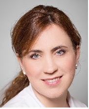
Maria Vasilyeva

Friendship University of Russia, Russia
Speaker

Co-Chair

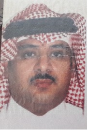
Mohammad Ali Albakry

Najran University, KSA

Session Introduction

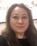
Ayça Dilara YILMAZ

Ankara University, Turkey

Title: Genetic Association Study Between Vitamin D Receptor Gene and Temporomandibular Disorders

Time : 10:00-10:30
Speaker
Biography:

Ayça Dilara YILMAZ has expertise in molecular biology and genetics. She has attended to different molecular biology departments working on hematology, pneumonia, asthma and epilepsy. She has done her master’s degree in Hematodiagnostics department of Ankara University Faculty of Medicine. She has completed her Phd in Ankara University Biotechnology Institute Proteomics Department in 2012. She has been working as a researher in the molecular biology laboratory of Dentisty Faculty since her graduate. She has been working with dentistry students and research fellows on many projects including rat models and zebra fish model. Research in periodontitis, maxillofacial genetics, PRP treatment for improving the health and wellbeing are her recent studies in the field.

Abstract:

Aim: Temporomandibular joint internal derangement (TMJ-ID) is the imbalance of metabolic processes in the extracellular matrix (ECM) of the articular disc, which progressively degrades causing tissue breakdown. The Vitamin D receptor (VDR) gene polymorphisms have been investigated for their potential effects and functional significance on several pathological conditions particularly osteoarthritis (OA) and disc degeneration-linked pathologies. The aim of this study was to investigate the possible association of Fok1, Apa1 and Taq1 polymorphisms of VDR gene with TMJ-ID.
Materials and methods: The study included 49 unrelated TMJ-ID patients (31.7 ±7.9) and 70 healthy controls (28.22 ±5.9) without TMJ-ID. Additionally, TMJ-ID patients were evaluated as anterior disc displacement with reduction (ADDWR) (n=24) and anterior disc displacement without reduction (ADDWOR) (n=25). Blood samples were obtained and DNA was extracted by standard proteinase K/phenol-chloroform method. Fok1, Apa1 and Taq1 polymorphisms of VDR gene were investigated by a polymerase chain reaction (PCR) based restriction fragment length polymorphism (RFLP).
Results: The genotype and allele frequency distributions of Fok1/rs2228570 (C>T), Taq1/rs731236 (T>C) and Apa1/ rs7975232 (A>C) did not show significant differences in TMJ-ID patients compared to the healthy group. In Fok1, carrying the TT genotype was almost 2 fold risk factor in TMJ-ID, ADDWR and ADDWOR patients compared to the healthy group (OR=1.72, OR=1.55, OR= 1.93 respectively) although not significant. In ADDWR, CT genotype was significantly different
than CC genotype (OR=0.35, CI:0.12-1.02, p<0.05) as a protective factor. In Apa1, carrying the AC and CC genotype was almost 1.23-1.79 fold risk factor in TMJ-ID patients, in ADDWR and ADDWOR cases compared to the healthy group although not significant. There were no significant results in none of the groups in Taq1 polymorphism.
Conclusion: Our results suggest that Fok1 and Apa1polymorphisms may be associated with TMJ-ID pathogenesis. Increasing the case and controls numbers is needed to further evaluate the genotype and allelic frequencies and risk factor ratios of VDR
polymorphisms in TMJ-ID.

Pedro Carvalho Feitosa

São Paulo University of São José dos Campos , Brazil

Title: The socket-shield technique to for Ridge Preservation at immediate implant placement

Time : 10:45-11:15

Speaker
Biography:

Pedro Carvalho Feitosa holds a degree in Dentistry from the University of Santo Amaro (1995) with specializations in Oral and Maxillofacial Surgery and Traumatology, Master’s in Implant Dentistry (UNISA), PhD in Implantology (São Leopoldo Mandic), and Biopathology from Universidade Paulista (UNESP), São José dos Campos - Sp. He is currently the Coordinator of the Institute of Dental Research IPEO - São Paulo (FACSETE). He is also the Coordinator for specialization course in Implantology at the University of Pindamonhagaba (FAPI) at GRAAL, São Luís-Ma. He is Professor of Implantology at CEOSP; University of Taubaté; Estácio de Sá campus São José dos Campos (CEDDA). He is the International Coordinator in Implantology at UNINORTE, and Professor of the specialization course in Maxillofacial Surgery and Traumatology of the São Leopoldo Mandic University in Campinas.

Abstract:

After extraction of the teeth, the alveolar bone goes through the remodeling process, which leads to horizontal and vertical bone loss. These reabsorption processes complicate dental rehabilitation, particularly in connection with implants. Careful management of post-extraction tissues is necessary to preserve the alveolar ridge. Several methods of guided bone regeneration have been described to retain the original bone size after extraction. Most procedures use filling materials and membranes to support the oral plaque and soft tissues, stabilize the clot and prevent epithelial growth. Instead of surgical augmentation to correct a defect peak, the socket-shield technique offers a promising solution. With this method, it will be demonstrated how the crest and the entire buccal bone are clinically preserved and the biological integrity of the buccal periodontium remains untouched. This method has also been described in connection with the immediate placement of the implant and the provisional crown. The immediate postoperative period, as well as the follow-up of one year, will be demonstrated as a functional and aesthetic result of the treatment. The socket-shell technique is a highly promising addition to clinical implant dentistry. We will discuss the possibilities, limitations, and guidelines for future research that we begin at our university.

Speaker
Biography:

Uri Zilberman received his DMD degree in 1983 from the Dental Faculty, Hebrew University, Jerusalem, and his PhD degree in 2000 in Basic Science, Dental Anthropology. He specialized in Pediatric Dentistry from 1990 and has been treating children and adolescents for the last 30 years. He is the Head of the Pediatric Dental Unit at Barzilai Medical University Center. He is the Senior Lecturer at the Faculty of Health Science, Ben-Gurion University of the Negev, Beersheba and at the Dental Faculty, Hebrew University, Jerusalem. He teaches courses on Dental Morphology for first year Dental students and Pathophysiology of the Oral Cavity for fourth year Pharmacology students. He has published more than 50 research papers and chapters in Pediatric Dentistry and Dental Anthropology. His main interests are new dental procedures and devices for pediatric dentistry, hereditary disorders and their effect on tooth development, and the use of biomimetic materials in pediatric dentistry, like glass-ionomers cements. He has developed new dental procedures and a patented new dental device for pediatric dentistry.

Abstract:

Missing upper anterior deciduous teeth may be caused by trauma or due to extraction as a result of extensive carious attack. The premature loss of anterior dentition may affect eating, speech development and behavioral problems. The parents' attitude toward restoration of damaged, unaesthetic or missing anterior teeth is very positive and may influence the final treatment. Several attempts to restore missing anterior teeth using polyethylene fibers were published with positive results. The use of glass fibers as a frame for restoration of missing permanent teeth showed success rate of more than 90% after 5-20 years. The use of glass fibers in pediatric dentistry poses several problems to the clinician: 1. the small size of the crowns that reduce the area of bonding. 2. the adhesive procedure which is affected by the thick a-prismatic layer of the deciduous teeth. 3. the length of the chair side procedure that is affected by the child age and tranquility. The aim of the study was to establish the optimal bonding procedure and bonding material for adhesion of glass fibers frame to deciduous teeth and to develop a method of preparing a preformed rigid frame for restoring missing upper anterior deciduous teeth. The presentation will show the results of the laboratory study and the clinical implication of the procedure with long follow-up periods.

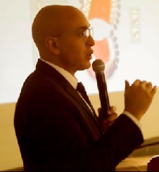
Abdelhalim Faris

Mansoura University, Egypt

Title: Materials Used in CAD/CAM Fixed Prosthodontics

Time : 12:30-13:00
Speaker
Biography:

Abdelhalim Faris is a Senior Technician, Faculty of Dentistry, Mansoura University, Egypt. He is the Founder of Elfaris Dental lab.

Abstract:

CAD/CAM dentistry is a field of dentistry and prosthodontics using computer-aided-design And computer-aidedmanufacturing to improve the design and creation of dental restorations, especially dental crowns, veneers, inlays and on lays, bridges, and dental implant restorations. There are a lot of materials available in the market to be used for different types of CAD/CAM Fixed prostheses. The aim of lecture is to discuss these different materials available in the market.

Biography:

Muhammad Aamir Ghafoor Chaudhary is an Assistant Professor at Department of Prosthodontics, Islamic International Dental College, Riphah International University, Islamabad, Pakistan.

Abstract:

The objective of this cross-sectional study was to determine the reliability of foveae palatinae for locating the position of the vibrating line in edentulous patients. The study was conducted in a tertiary care dental hospital in Islamabad, Pakistan on a sample of 150 completely edentulous patients. Clinical examination of the soft palate area was conducted to assess for the location of the vibrating line, whether it was at, anterior or posterior to the foveae. Interestingly, foveae were not present in 27.2% of the sample. 32.5% patients had foveae present at the level of vibrating line, 31.1% had vibrating line posterior to foveae palatinae, while only 8.6% presented with vibrating line anterior to fovea palatinae. Within the limitations of the present study, it was concluded that foveae palatinae are not a reliable anatomical landmark for determining the position of the vibrating line.

Hatim Barrak A AlOsaimi

Prince Sattam bin Abdulaziz University "PSAU", KSA

Title: Hatim's Matrix Band

Time : 16:15-16:45
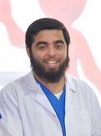
Speaker
Biography:

Hatim Barrak A.AlOsaimi completed his BDS from Prince Sattam bin Abdulaziz University "PSAU", Alkharj, KSA with GPA is 3.85 out of 5.00. Work with Sattam Bin Abdulaziz University, AlKharj, KSA - 2015, June - 2015,Oct also join as an intern at Prince AbdulRahman Advanced Dental institute , Riyadh, KSA- Oct, 2015 – Feb 2016. He is a Head of 7th Dental Conference At Prince Sattam Bin Abdulaziz University- 2016. He is an Organizer in Community Dental Program ( MUSHREQAH I ) At Alkharj City – 2013.

Abstract:

From being one of the dentists; I have been thinking for creating an instrument to help in restoring the most difficult types of dental caries, (CLASS II Cavity), because it is a caries related to the teeth adjacent to it, which make it difficult to restore it and for that reason we are using an instrument in restoring this type of caries. So in which that instrument be easier, flexible and integrated than what is now available in dental clinics. Therefore, when looking to the instruments which are using in dental clinics now-a-days for restoring (CLASS II CAVITY), we find dis-advantages including the following: Most of it does not take the form of tooth curvature or not formed on the form that required to make the filling compensates of caries of the tooth properly and intimate fit to the composition of the tooth, which prevents future caries due to lack or increase the filling of the caries cavity as well as do not maintain the distance between teeth, making it difficult in using of dental floss and so on. And most of these instruments cause injury and damage gingival structure due to a thickness of the unit of these instruments and its types are completely incompatible with the gingival pockets, which is necessary to allowing entering these instruments, especially if the caries is under CEJ. The innovative instrument has the following characteristics: It has a strong ability to form around the tooth, which gives a great ability to take the form of filling cavity only, which makes it easy to keep the distance between the teeth to be cleaned and thus prevent the occurrence of any subsequent caries. And does not cause any injuries to the gingiva because it is too thin and sharp so easily flow it in the pockets of gingiva, especially When decay is below the CEJ line .And also the instrument is hydrophobic, which helps to prevent the arrival of liquids to the cavity and this is important for the success of filling. Marketing quality is possible because this instrument is disposable material and very symbolic value also I has been formed a good number of them and use it in phantom, which proved the feasibility of the idea and its effectiveness.